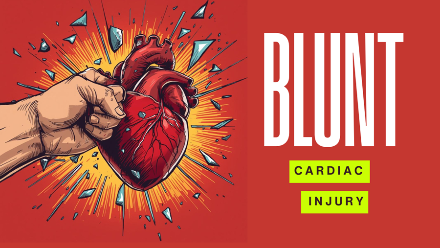
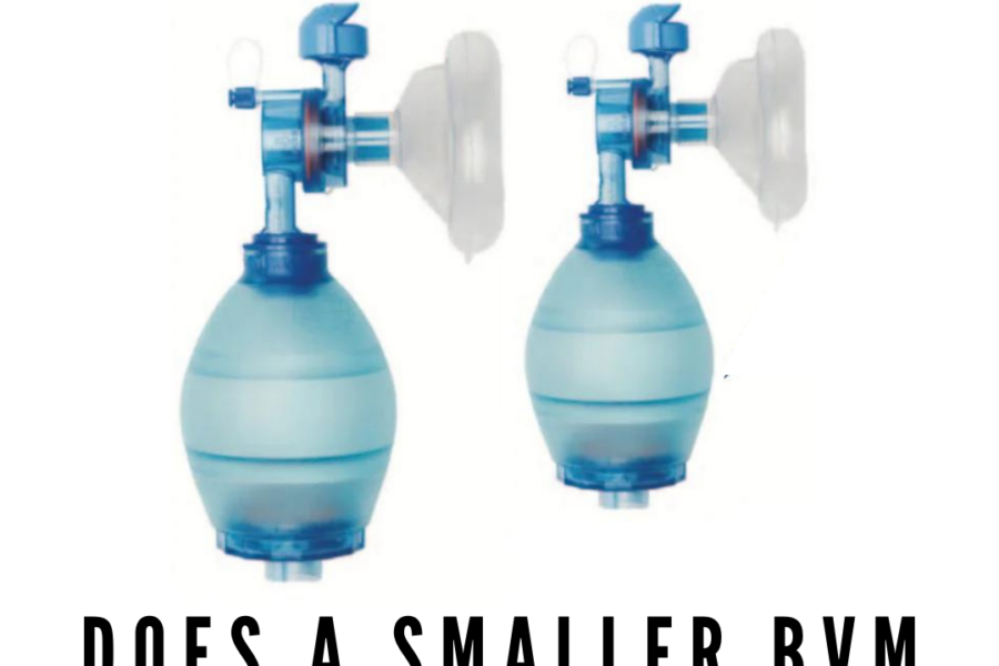
BlogEMCORE Newsletter Blunt Cardiac Injury BLUNT CARDIAC INJURY What does the Evidence say? ⚡ Clinical Takeaways Isolated sternal fractures…Peter Kas7 March 2026
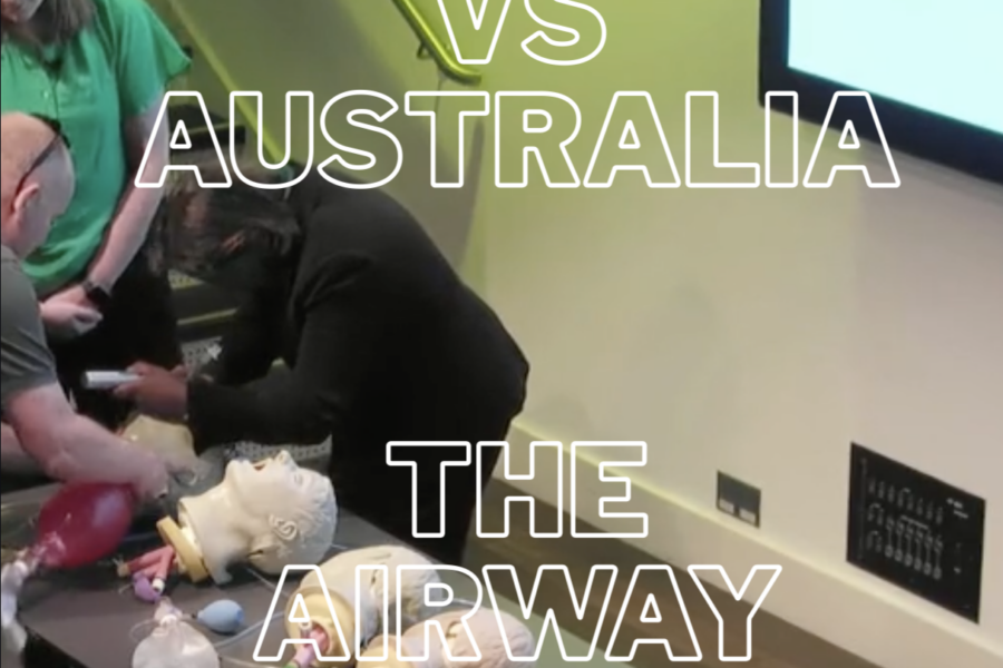
BlogEMCORE NewsletterUncategorised Airway Ashes Australia Wins the Cup! A short fun video that shows the Airway Ashes: England vs…Peter Kas27 January 2026
BlogEMCORE NewsletterUncategorised How EM Experts Think This week we feature Dr Reuben Strayer, who is speaking at EMCORE Singapore 15th -…Peter Kas7 January 2026
Uncategorised EMCORE Fiji 2025 Wrapped EMCORE Fiji 2025 Wrapped September saw us return to one of our favourite places on…Tessa Milton4 December 2025
EMCORE Newsletter EMQA 23/4/25: Learnings from the Latest EMCORE Conference EMCORE Sydney 2025 was a great conference full of learning. Here are just a few…Peter Kas23 April 2025
EMCORE Newsletter Resuscitation Immersion & Expertise You can't be an expert at Resuscitation and take yourself to the next level..... .....if…Peter Kas17 March 2024
EMCORE NewsletterEMQ&A EMQA 08/03/24 EMQA 08/03/24 Here's what I've got for you this week: A 40 yo with depressed…Peter Kas8 March 2024
EMCORE NewsletterEMQ&A EMQA 01/03/24 EMQA 01/03/24 Thank you to everyone who has asked that the EMQA format be brought…Peter Kas1 March 2024
EMCORE Newsletter EMCORE Newsletter 23/02/24 EMCORE London and Fiji are filling fast.The accommodation link for RESUSCORE in Bali, is now…Peter Kas23 February 2024
EMCORE Newsletter EMCORE Newsletter 15/02/24 EMCORE Newsletter 15/02/24 This week I'm back after a good break and ready for some…Peter Kas15 February 2024