

EMQA 08/03/24
Here’s what I’ve got for you this week:
- A 40 yo with depressed conscious state and fever and a history of IVDU, has an ECG that reads as “Acute AMI”. Is it? What else could it be?
- A Free 7 minute Video for you to watch: “Is My Patient Going to Die?”
- On The Shoulders of Giants: A short look at the amazing life of Dr Henning Ruben, anaesthetist, dancer, magician, who gave us amongst other things…the BVM.
- RESUSCORE Bali is going to be incredible! Seats are moving fast. Book Now.
- The Best of EMCORE Conference Lectures available now. 18 Lectures, with Certificate on completion.
E: ECG of The Week
A 40 yo is brought to the emergency department by ambulance, following being found in a chair, by a friend and could not be woken. There is a history of IVDU. There is no sign of injury and the vitals are:
GCS 4, SBP 70mmHg, HR 140, RR 50, T 39C
Heart sounds are dual with no extra sounds. There are reduced breath sounds and crepitations in the left base. The abdomen is soft and the patient appears to have peripheral cyanosis.
You prepare for intubation. Fluids and inotropes and antibiotics are given and an ECG is done.
What does the ECG show?
What are your differential diagnoses for this patient? What is the most likely diagnosis?
Let’s look at the ECG, then click below for the ECG analysis.

**CLICK HERE to see ECG Description**
Rate 106
Is it sinus?– P waves are upright in II and inverted in aVR; Yes
Is there a P for every QRS? Yes
QRS:
Is it tall/small?: It’s about right, there is no hypertrophy.
Is it wide/narrow? It’s narrow
Is it of abnormal morphology ie., delta wave? No
Is it clumped?(just incase we miss a Mobitz- but it’s not slow enough) No
ST-T
Remember the baseline is the T-P line
If we look we see that there is some ST elevation in some areas of the ECG of about 1mm, although this is difficult to gauge as the T wave goes straight into the p wave.
The most visible abnormality is the inverted T waves.
What causes inverted T waves? Causes include:
- Acute Myocardial Infarction
- Ventricular Hypertrophy( not present on this ECG)
- Pulmonary Embolism
- Raised Intracranial Pressure( I would usually expect them to be deeper)
PR and QT Intervals
The PR is normal and the QT is very prolonged.
PACING Spikes: None.
Putting it all together, what would seem the most probable diagnosis?
One differential to entertain is an overdose, however there are no signs of classic QRS widening of Na channel blockade, or a terminal R wave in aVR.
It might be an AMI, there is no evidence of hypertrophy, the patient is not behaving like they have a PE, but they may have raised intracranial pressure given the decreased conscious state. The decreased conscious state may be simply due to low blood pressure or an overdose.
The patient is also febrile. Given the history of IVDU, infective endocarditis is a consideration. The patient may also have meningitis. The fever may also be secondary to stroke as we know that about 50% of stroke develop a fever, which in itself is a marker of severity and marker of mortality.
Let’s narrow it down to 2 causes, AMI and raised intracranial pressure. Raised intracranial pressure ECG’s have deep T wave inversion and a prolonged QT. I’m thinking this is an intracranial event, especially if the fever is due to this.
Echo showed a poorly contracting myocardium, with clean valves, so it may be AMI.
ECG abnormalities if they don’t make sense may not be coming from the heart.
The CT gave the diagnosis with large subarachnoid haemorrhage.
This is a neurocardiogenic phenomenon when the heart is affected by deranged autonomic supply coming from a stroke. A fascinating case.
Free Video for the Week: “Is my Patient going to die?”
Dr Pascal Galperowics, gives us a quick review: It’s from the Vault.
Q: Quote
“I saw the angel in the marble and carved until I set him free”
Michaelangelo Buonarroti
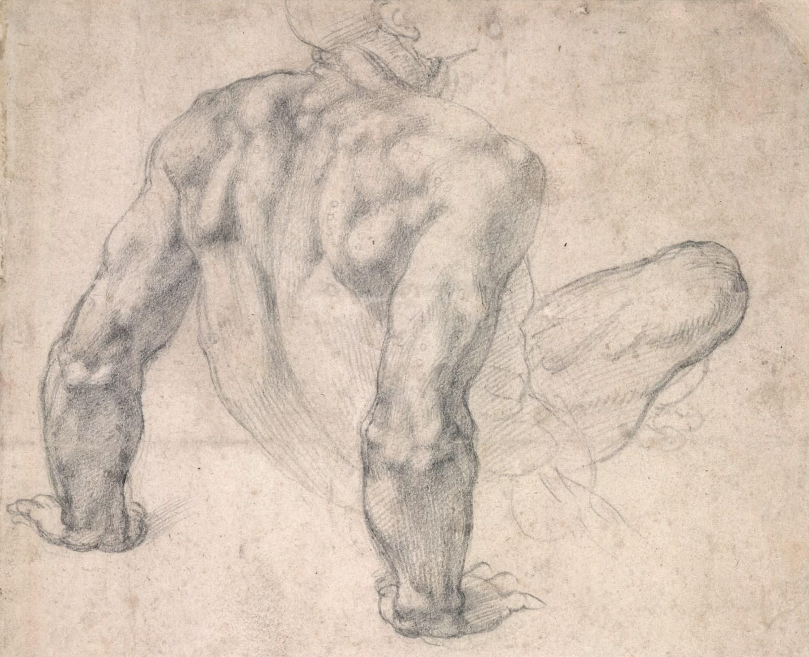
Art: Because It’s not all about Medicine.
On the Shoulders of Giants
Dr Henning Ruben (1914-2004)

- At the age of 19 in 1933, he enters the school of dentistry in Copenhagen. He studies dentistry but that’s not all.
- He is also a professional dances. His speciality: The tango.
- He is a member of the Danish Fencing team and wins a Bronze Medal at the World Fencing Championships in 1939
- He is also an accomplished magician: A mind reader.
In 1943 he enters medical school. Unfortunately the Nazi occupation of Denmark and his Jewish ancestry requires him to flee, which he does so in the dead of night on a Fishing boat bound for Sweden, where he stayed for 2 years.
In Stockholm he worked as a dentist and as a magician. Whilst there, he becomes interested in anaesthesia, which is far more advanced as a speciality in Sweded, than in Denmark. However he returns to Copenhagen after the war and graduates.
His desire to travel and study is great but travel was difficult. Sometimes fate intervenes. In 1947 he is invited to Sweden by the Swedish Society of Illusionists. Whist there he meets wth hospital anaethetists and the following year, he is appointed at Sabbatsberg and St Ericks hospital.
His inventions include:
- 1948- He developed the Ruben Valve a non-rebreathing Valve
- 1953- Invents the foot-operated sucker.
- 1954- He developed a constant rate syringe pump driven by a wind-up alarm clock for drug delivery
- 1954- He invents the self-inflating bag valve mask
…… I’ll speak more on Dr Ruben at EMCORE London.
SEE YOU AT EMCORE
Less than 30 places left for London.


JOIN US FOR OUR OTHER COURSES
The Cardiac Online Course
Primary Exam Course
Written Fellowship Course
The BRAND NEW OSCE Course
JOIN US at Cardiac Bootcamp Pre-Conference Workshop
COMPLETED BY THOUSANDS OF DOCTORS, NURSES AND PARAMEDICS

Thankyou always for all your hard work.
“The knowledge you take into your shift DOES matter” Peter Kas

Due to the volume of emails from thousands of weekly readers, I am unable to read and reply to them all.
Forward this to someone.
Received this from someone? Join the thousands of weekly readers JOIN NOW
Keep in touch with what’s happening at the emcore conferences by joining us on social media @emcoreshow.